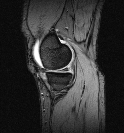
MRI of Knee showing a tear of the Medial Meniscus.The meniscus has a triangular appearance which is changed when the structure is damaged – split horizontally here.

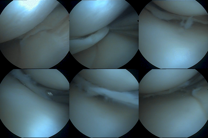
Meniscal tear- Unstable cartilage pieces pull on lining of the joint causing pain and mechanical symptoms such as locking or catching.
Arthroscopy For Cartilage Damage
What is Knee Arthroscopy?
Knee arthroscopy is a common surgical procedure in which the knee joint is viewed using a small high definition camera. This allows the diagnosis and treatment of a range of knee conditions.
What does Knee arthroscopy involve?
Arthroscopy is done through small incisions (keyhole surgery).
During the procedure a small telescope about the size of a pencil is inserted into your knee joint. The high definition camera attached to this arthroscope sends the image to a high resolution television monitor.
Structures of the knee can be seen and tested in great detail.
Using small specialised surgical instruments inserted through other incisions around your knee Mr Jennings can use arthroscopy to feel, repair or remove damaged tissue.
The surgery usually takes between 30 and 60 minutes to complete and is carried out under general anaesthetic as a day case procedure. Prior to your surgery details of your treatment and recovery will be discussed with you by Mr Jennings.
What is a torn cartilage?
There are two meniscal cartilages in the knee. The inner (medial) or outer (lateral) meniscus. These are made of specialised collagen fibres and act to spread the load through the knee, as a sort of shock absorber.
The meniscus can be torn or damaged leading to mechanical irritation of the knee. The cartilage itself has no nerves in it but it is attached to the lining of the knee which is very sensitive. If the damaged cartilage pulls on the sensitive lining it will cause pain. Occasionally big tears may cause mechanical problems, such as giving way and lack of extension (when the knee cannot go straight or is “locked).
Treatment can be either through pain relief and rehabilitation or by surgery to either repair the cartilage or remove the damaged parts causing the irritation. All treatment options will be discussed in full with you. Often the information from an MRI scan can help steer you to the best management plan to suit your symptoms and needs. Repair of the cartilage often requires a slower recovery and protecting the cartilage in a knee brace for several weeks.
Recovery
Your recovery program will start immediately and will be tailored to your surgery and requirements. You will be instructed on basic self-directed exercises for the immediate post-operative period. In most cases you will walk out of the hospital.
Keep wounds clean, dry, and covered at all times until outpatient review. The waterproof dressing will allow showering but not bathing/immersion.
Mr Jennings will go through details of your surgery before your discharge and will go over this again when you return to the out-patient clinic for review approximately 2 week later.

MRI of Knee showing a tear of the Medial Meniscus.The meniscus has a triangular appearance which is changed when the structure is damaged – split horizontally here.

Meniscal tear- Unstable cartilage pieces pull on lining of the joint causing pain and mechanical symptoms such as locking or catching.


